重组抗体
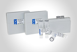
重组抗体,也称为基因工程抗体,是指通过DNA重组技术将抗体相应的基因序列根据需要进行改造和重组,并构建在质粒上,再通过蛋白外源表达技术将构建好的质粒转染/转化入适合的宿主细胞表达获得的抗体。换言之,重组抗体是通过将免疫特异性重链和轻链抗体克隆到高产的哺乳动物表达载体中而制成的。将得到的载体引入到表达宿主(如细菌、酵母或哺乳动物)中,用于生产高质量的功能性抗体。重组抗体很好的解决了动物源抗体引起的人体排斥反应,使得抗体实现人源化,使抗体的效能更为完善。
重组抗体可分为五大类:嵌合抗体、人源化抗体、全人源化抗体、小分子抗体、双特异性抗体。近年来,重组抗体的使用在治疗和诊断中变得越来越广泛。与传统抗体相比具有显著优势,卓越的批次间一致性、持续供应和非动物源优势使得重组抗体变得越来越受青睐,最重要的一点是重组抗体可以最大化人源化以解决不同物种之间的异质性。
j9九游会登录入口首页生物目前已开发1000多种重组抗体,具有高纯度、亲和力强以及批次间偏差小等特点,涉及癌症、心血管疾病、细胞生物学、免疫学、神经科学、表观遗传学和核信号传导、信号转导等众多领域。为了更好地帮助客户进行科学研究,CUSABIO还成功生产了针对许多困难靶点的重组抗体,如:CLDN18.2、CLDN6、CCR8、SSTR2、DLL3、LY6G6D、ROR1、SEMA4D、GPC3、CD16A等。
产品优势
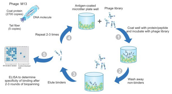
● 先进的噬菌体展示技术
j9九游会登录入口首页生物一直专注于将先进的噬菌体展示技术应用于广泛的服务项目,特别是重组抗体的开发。
● 重组抗体种类繁多
j9九游会登录入口首页生物可提供各种类型的重组抗体,包括全长抗体、scFv、Fab、sdAb、同种型/亚型和各种形式的 Fc 融合蛋白。

● 多种应用类型可供选择
j9九游会登录入口首页生物的重组抗体已在多个应用平台验证,包括 WB、IF、IHC、FC、IP、ELISA、GICA 和 Neutralising。

Phospho-ERN1 (S724) Antibody (CSB-RA007795A724phHU)
Positive WB detected in: 293 whole cell lysate, A549 whole cell lysate, Hela whole cell lysate (treated with Calyculin A or EGF)
All lanes: Phospho-ERN1 antibody at 0.75μg/ml
Secondary
Goat polyclonal to rabbit IgG at 1/50000 dilution
Predicted band size: 110 kDa
Observed band size: 110 kDa
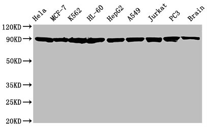
HSP90AA1/HSP90AB1 Antibody (CSB-RA010802A0HU)
Positive WB detected in: Hela whole cell lysate, MCF-7 whole cell lysate, K562 whole cell lysate, HL-60 whole cell lysate, HepG2 whole cell lysate, A549 whole cell lysate, Jurkat whole cell lysate, PC3 whole cell lysate, Rat brain tissue
All lanes: Hsp90 alpha + beta antibody at 1.25μg/ml
Secondary
Goat polyclonal to rabbit IgG at 1/50000 dilution
Predicted band size: 90 KDa
Observed band size: 90 KDa
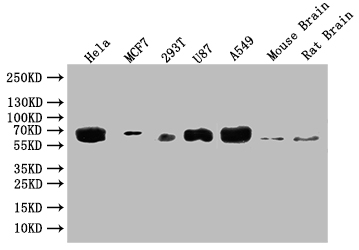
GBA Antibody (CSB-RA869334A0HU)
Positive WB detected in: Hela whole cell lysate, MCF7 whole cell lysate, 293T whole cell lysate, U87 whole cell lysate, A549 whole cell lysate, Mouse Brain tissue, Rat Brain tissue
All lanes: GBA antibody at 1:500
Secondary
Goat polyclonal to rabbit IgG at 1/50000 dilution
Predicted band size: 60 kDa
Observed band size: 60 kDa

VDAC1 Antibody (CSB-RA025821A0HU)
Positive WB detected in: Hela whole cell lysate, HepG2 whole cell lysate, 293 whole cell lysate, Jurkat whole cell lysate, HL-60 whole cell lysate, LO2 whole cell lysate, Raji whole cell lysate, Rat heart tissue, Mouse brain tissue
All lanes: VDAC1 antibody at 0.7μg/ml
Secondary
Goat polyclonal to rabbit IgG at 1/50000 dilution
Predicted band size: 31 KDa
Observed band size: 31 KDa
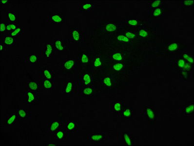
Histone H3.3 Antibody (CSB-RA010109A0HU)
Immunofluorescence staining of Hela cells with the antibody at 1:60, counter-stained with DAPI.
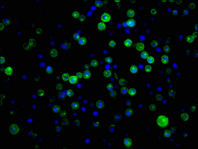
CD21 Antibody (CSB-RA005934A0HU)
Immunofluorescence staining of Raji cells with the antibody at 1:34, counter-stained with DAPI.
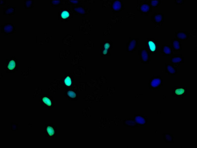
Histone H3.1 Antibody (CSB-RA010418A0HU)
Immunofluorescence staining of Hela cells with the antibody at 1:93, counter-stained with DAPI.
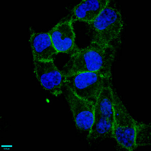
CD97 Antibody (CSB-RA004972A0HU)
Immunofluorescence staining of Hela cells with the antibody at 1:87.5, counter-stained with DAPI.
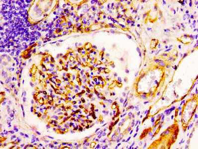
CD34 Antibody (CSB-RA004926A0HU)
IHC image of the antibody diluted at 1:100 and staining in paraffin-embedded human kidney tissue performed on a Leica BondTM system.
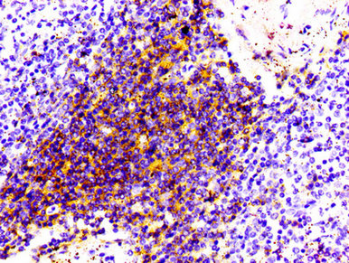
CD4 Antibody (CSB-RA004935A0HU)
IHC image of the antibody diluted at 1:100 and staining in paraffin-embedded human spleen tissue performed on a Leica BondTM system.
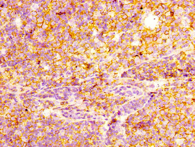
CD44 Antibody (CSB-RA004938A0HU)
IHC image of the antibody diluted at 1:100 and staining in paraffin-embedded human tonsil tissue performed on a Leica BondTM system.
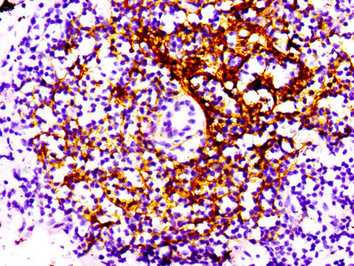
CD21 Antibody (CSB-RA005934A0HU)
IHC image of the antibody diluted at 1:100 and staining in paraffin-embedded human lung cancer performed on a Leica BondTM system.
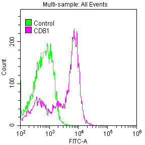
CD81 Antibody (CSB-RA004960A0HU)
Overlay histogram showing Jurkat cells stained with the antibody (red line) at 1:50. Control antibody (green line) was used under the same conditions. Acquisition of >10,000 events was performed.
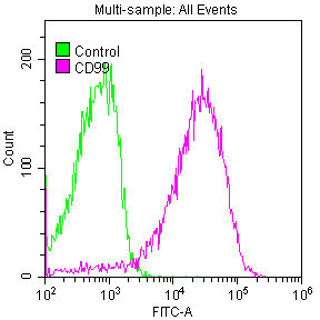
CD99 Antibody (CSB-RA004973A0HU)
Overlay histogram showing Jurkat cells stained with the antibody (red line) at 1:50. Control antibody (green line) was used under the same conditions. Acquisition of >10,000 events was performed.
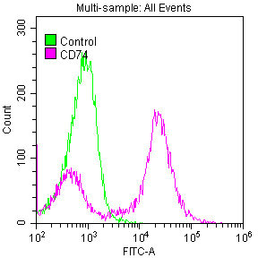
CD74 Antibody (CSB-RA004956A0HU)
Overlay histogram showing Raji cells stained with the antibody (red line) at 1:50. Control antibody (green line) was used under the same conditions. Acquisition of >10,000 events was performed.
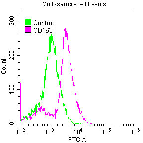
CD163 Antibody (CSB-RA801238A0HU)
Overlay histogram showing Raw264.7 cells stained with the antibody (red line) at 1:50. Control antibody (green line) was used under the same conditions. Acquisition of >10,000 events was performed.

Phospho-CDK2 (Y15) Antibody (CSB-RA005061A15phHU)
Positive WB detected in:Hela whole cell lysate, 293 whole cell lysate(treated with Pervanadate or not)
All lanes: Phospho-CDK2 antibody at 0.8μg/ml
Secondary
Goat polyclonal to rabbit IgG at 1/50000 dilution
Predicted band size: 34 KDa
Observed band size: 34 KDa
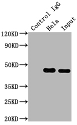
MAP2K1 Antibody (CSB-RA957619A0HU)
Positive WB detected in: Hela whole cell lysate, 293 whole cell lysate, MCF-7 whole cell lysate, 293T whole cell lysate, A549 whole cell lysate, U251 whole cell lysate, Rat brain tissue
All lanes: MAP2K1 antibody at 1:2000
Secondary
Goat polyclonal to rabbit IgG at 1/50000 dilution
Predicted band size: 44, 41 kDa
Observed band size: 44 kDa
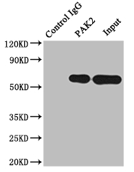
PAK2 Antibody (CSB-RA592787A0HU)
Positive WB detected in: 293 whole cell lysate, Jurkat whole cell lysate, Raji whole cell lysate, Mouse brain tissue, Rat brain tissue
All lanes: PAK2 antibody at 1:2000
Secondary
Goat polyclonal to rabbit IgG at 1/50000 dilution
Predicted band size: 59 KDa
Observed band size: 59 KDa
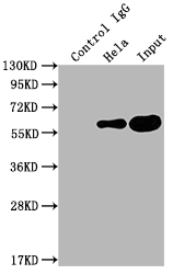
PKM Antibody (CSB-RA632595A0HU)
Positive WB detected in: SH-SY5Y whole cell lysate, Jurkat whole cell lysate, MCF-7 whole cell lysate, Hela whole cell lysate, Raji whole cell lysate, 293 whole cell lysate, Mouse brain tissue
All lanes: PKM antibody at 1:2000
Secondary
Goat polyclonal to rabbit IgG at 1/50000 dilution
Predicted band size: 58, 59, 57 kDa
Observed band size: 58 kDa
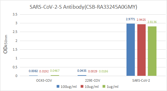
Immobilize various types of SARS proteins at concentration of 2μg/ml on solid substrate, then react with SARS-CoV-2-S Antibody at concentration of 100μg/ml, 10μg/ml and 1μg/ml. It shows the SARS-CoV-2-S Antibody (CSB-RA33245A0GMY) is specific for SARS-CoV-2-S1-RBD protein, without any cross-reactivity with HCoV-OC43, HCoV-229E.
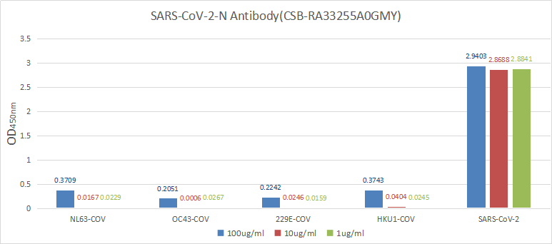
Immobilize various types of SARS proteins at concentration of 2μg/ml on solid substrate, then react with SARS-CoV-2-N Antibody at concentration of 100μg/ml, 10μg/ml and 1μg/ml. It shows the SARS-CoV-2-N Antibody (CSB-RA33255A0GMY) is specific for SARS-CoV-2-N protein, without any cross-reactivity with NL63-CoV, HCoV-OC43, HCoV-229E or HCoV-HKU1.
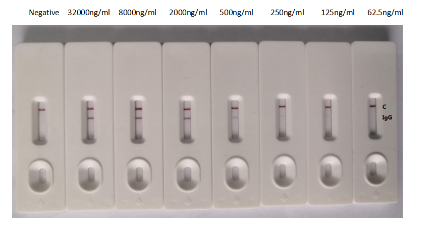
In the Colloidal Gold Immunochromatography Assay detection system, the background of antibody (CSB-RA33255A1GMY) is clean, the detection limit can be as low as 125ng/mL (8.75ng/0.07mL), and the sensitivity is very good.
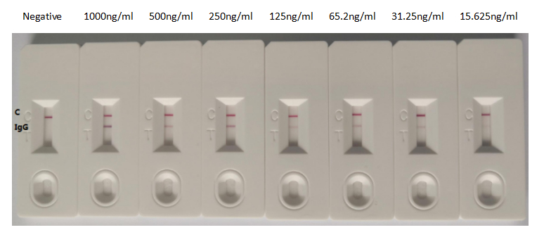
In the Colloidal Gold Immunochromatography Assay detection system, the background of antibody (CSB-RA33255A2GMY) is clean, the detection limit can be as low as 15.625ng/ml (1.09ng/0.07ml), and the sensitivity is very good
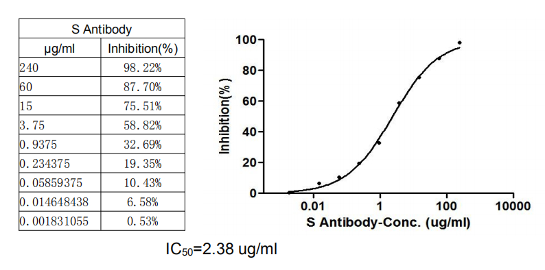
Binding signal of SARS-CoV-2-S1-RBD (CSB-YP3324GMY1) and ACE2 protein-HRP conjugate (CSB-MP866317HU) was inhibited by S Antibody (CSB-RA33245A1GMY) with the IC50 is 2.38 μg/Ml.
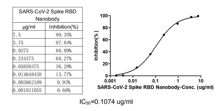
SARS-CoV-2 Spike RBD Nanobody (CSB-RA33245A2GMY)
This nanobody competitively prevented SARS-CoV-2-S1-RBD (CSB-YP3324GMY1) from binding to ACE2-HRP conjugate (CSB-MP866317HU). The inhibition efficacy of the SARS-CoV-2-S1-RBD/ACE2 binding was positively proportionally to the nanobody concentrations. It showed that the nanobody effectively inhibited the SARS-CoV-2-S1-RBD/ACE2 binding. And the IC50 of the nanobody is 0.1074 μg/mL.
● 批间差异小
● 可持续性供应
产品列表
您可以在下述列表中寻找您所需要的产品,或直接在顶部搜索框中输入您需要查找的靶点、产品名称、货号。有任何问题,请点击此处在线咨询。
关于重组抗体
与传统抗体相比,重组抗体的优势具体如下:
- 更高的一致性和重复性
因为重组抗体是由一组独特的基因发展而来的,所以抗体生产是可控且可靠的。可以避免杂交瘤细胞制备中的一些问题,如基因丢失、基因突变和细胞株漂移等。这使得抗体的批次间差异非常小,从而为您提供高度可重复的结果。
- 更高的灵敏度和特异性
利用重组技术,更容易通过抗体工程提高抗体特异性和灵敏度。所需克隆的选择过程发生在杂交瘤细胞和重组克隆阶段,这使我们能够选择最优的抗体质量。
- 易于扩展
随着抗体基因的分离,与传统单克隆技术相比,抗体表达能够以任何规模在更短的时间内进行。这意味着我们可以在数周而非数月内产生定制抗体。
- 无动物高通量生产
一旦分离出产生抗体的基因,就可以实现无动物体外生产。
重组抗体与传统抗体的比较:
| 单克隆抗体 | 多克隆抗体 | 重组抗体 | |
|---|---|---|---|
| 重复性 | 高;批次间差异小 | 低 | 更高;高纯度,批次间差异更小 |
| 特异性 | 强 | 弱 | 更强 |
| 生产周期 | 4到6个月的开发;4到6周的生产 | 2到3个月的开发和生产 | 4个月的开发;1到6周的生产 |
| 同型亚基转换 | 困难 | 困难 | 方便 |
| 表达系统 | 小鼠腹水法和体外方法 | 通常在哺乳动物细胞(如兔) | 通常在哺乳动物细胞系中,也可在特殊工程细胞系中表达,如酵母、细菌、昆虫和转基因植物 |
| 成本 | 生产成本较高;需要更专业的人员操作 | 成本相对较低 | 需要技术专长和较大的时间和金钱投入进行生产 |
| 人类免疫应答 | 无法避免 | 无法避免 | 可避免 |
引用文献
S Antibody referenced in "A magneto-optical biochip for rapid assay based on the Cotton–Mouton effect of γ-Fe2O3@ Au core/shell nanoparticles", Journal of nanobiotechnology, 2021.
SARS-CoV-2 Spike RBD Nanobody referenced in "Potent antiviral activity of Agrimonia pilosa, Galla rhois, and their components against SARS-CoV-2", Bioorganic & Medicinal Chemistry, 2021.
HIF1A Antibody, CASP3 Antibody referenced in "PESV represses non-small cell lung cancer cell malignancy through circ_0016760 under hypoxia", Cancer cell international, 2021.
Phospho-ERN1 (S724) Antibody referenced in "The ER stress sensor inositol-requiring enzyme 1α in Kupffer cells promotes hepatic ischemia-reperfusion injury", Journal of Biological Chemistry, 2021.
CDK4 Antibody and CDK6 Antibody referenced in "Histone Deacetylase Inhibitor-Induced CDKN2B and CDKN2D Contribute to G2/M Cell Cycle Arrest Incurred by Oxidative Stress in Hepatocellular Carcinoma Cells Via Forkhead Box M1 Suppression", Journal of Cancer, 2021.
BIRC5 Antibody referenced in "LTBP4 Inhibits the Proliferation and Metastasis in Melanoma by Activating Hippo-YAP Signaling", Research Square, 2020.


