GFAP Recombinant Monoclonal Antibody
-
货号:CSB-RA009369MA1HU
-
规格:¥1320
-
图片:
-
Western Blot
Positive WB detected in: MCF7 whole cell lysate,Mouse Brain tissue lysate,Rat Brain tissue lysate
All lanes: GFAP antibody at 1:1000
Secondary
Goat polyclonal to mouse IgG at 1/50000 dilution
Predicted band size: 50 kDa
Observed band size: 55 kDa -
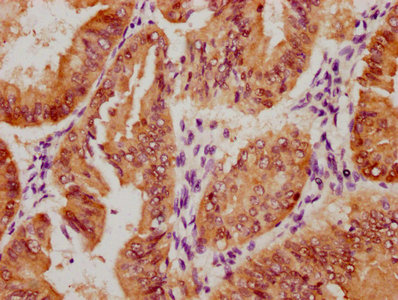
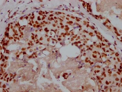
IHC image of CSB-RA009369MA1HU diluted at 1:300 and staining in paraffin-embedded human brain tissue performed on a Leica BondTM system. After dewaxing and hydration, antigen retrieval was mediated by high pressure in a citrate buffer (pH 6.0). Section was blocked with 10% normal goat serum 30min at RT. Then primary antibody (1% BSA) was incubated at 4°C overnight. The primary is detected by a Goat anti-Mouse IgG labeled by HRP and visualized using 0.05% DAB.
-
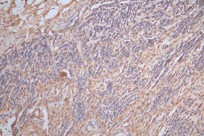
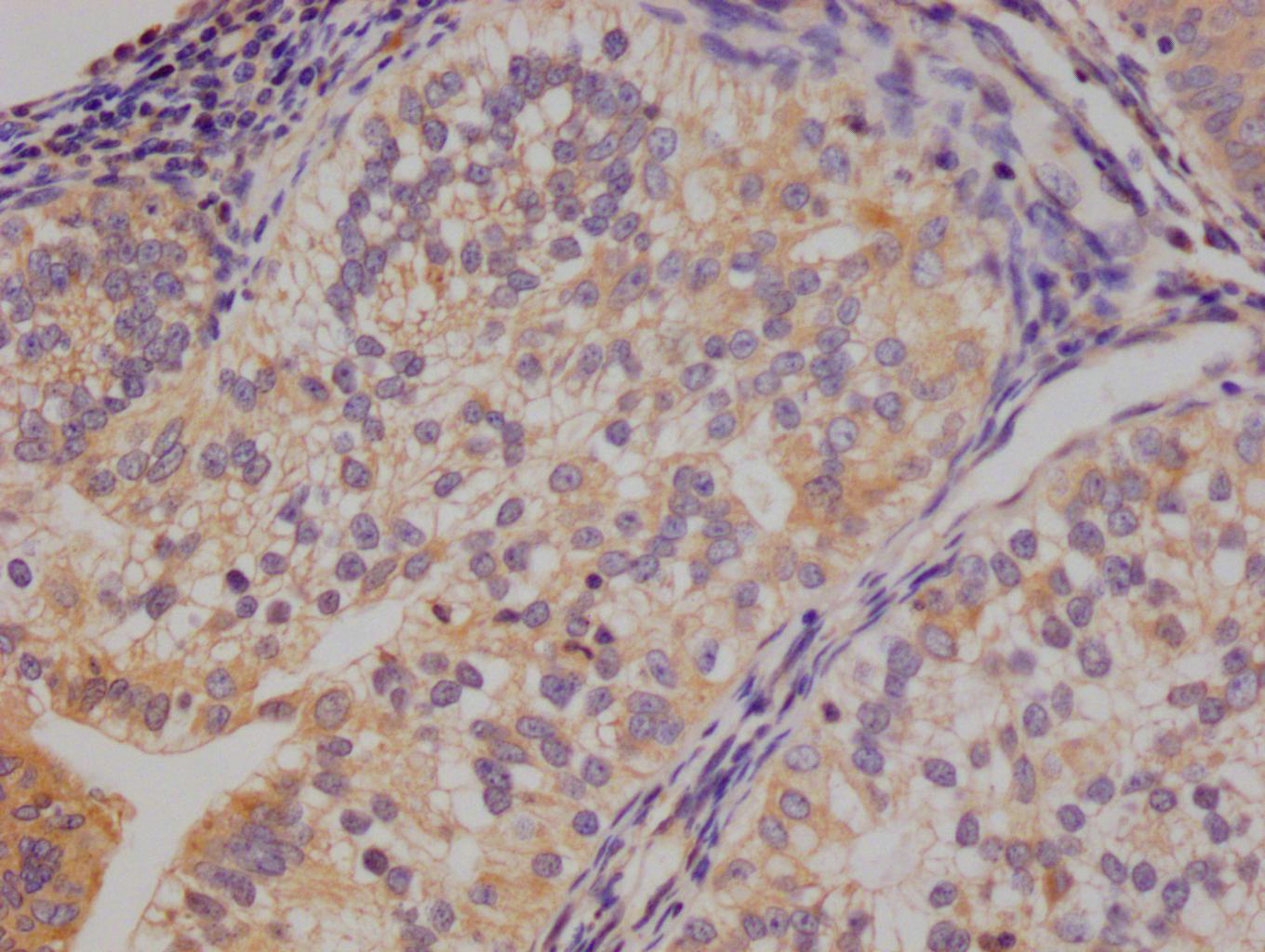
IHC image of CSB-RA009369MA1HU diluted at 1:300 and staining in paraffin-embedded human glioma cancer performed on a Leica BondTM system. After dewaxing and hydration, antigen retrieval was mediated by high pressure in a citrate buffer (pH 6.0). Section was blocked with 10% normal goat serum 30min at RT. Then primary antibody (1% BSA) was incubated at 4°C overnight. The primary is detected by a Goat anti-Mouse IgG labeled by HRP and visualized using 0.05% DAB.
-
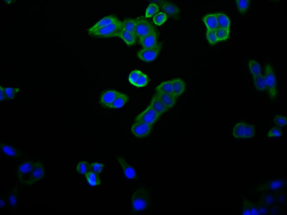
Immunofluorescence staining of SH-SY5Y cell with CSB-RA009369MA1HU at 1:200, counter-stained with DAPI. The cells were fixed in 4% formaldehyde and blocked in 10% normal Goat Serum. The cells were then incubated with the antibody overnight at 4C. The secondary antibody was FITC-conjugated AffiniPure Goat Anti-Mouse IgG(H+L).
-
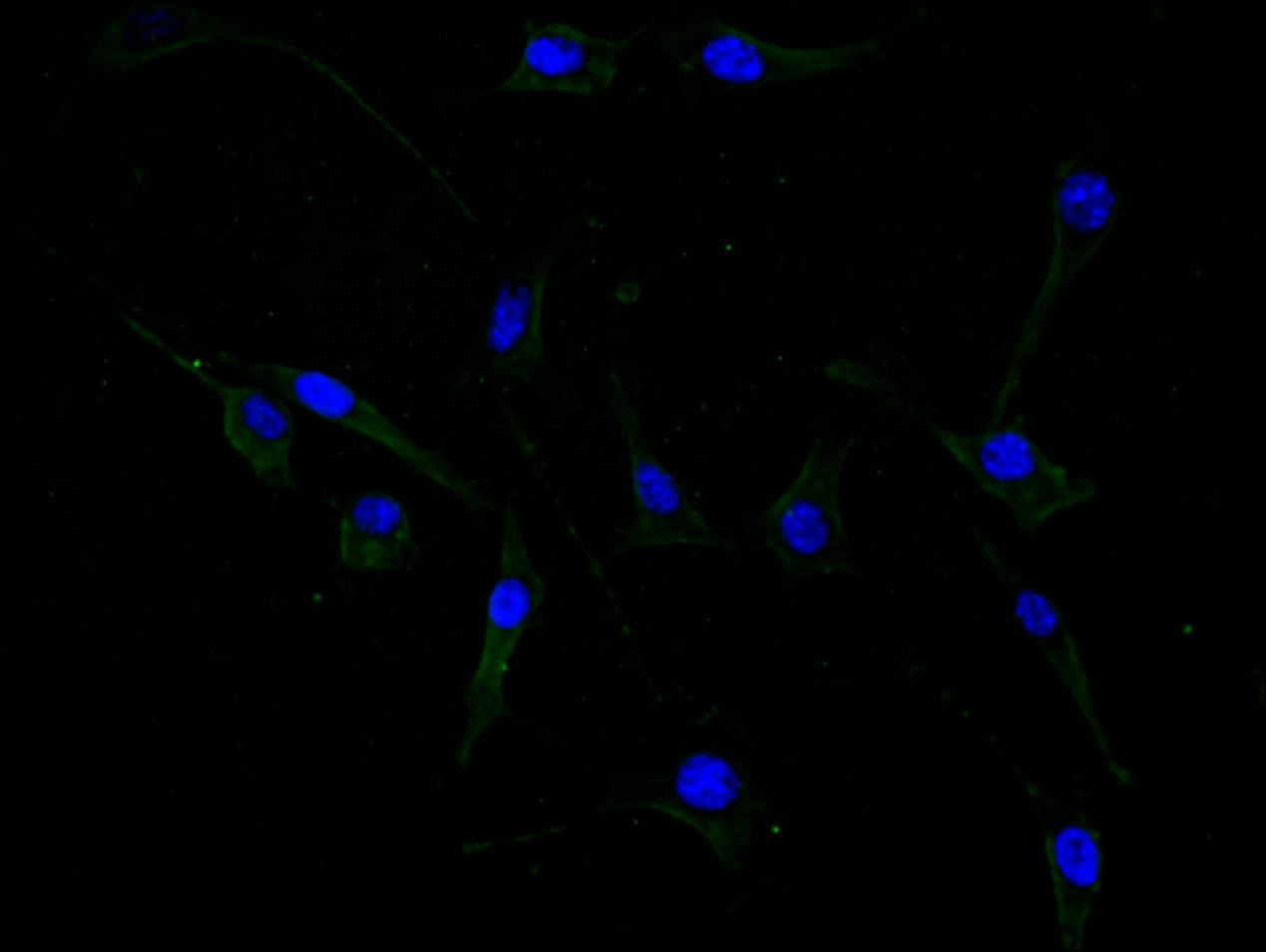
Immunofluorescence staining of U87 cell with CSB-RA009369MA1HU at 1:200, counter-stained with DAPI. The cells were fixed in 4% formaldehyde and blocked in 10% normal Goat Serum. The cells were then incubated with the antibody overnight at 4C. The secondary antibody was FITC-conjugated AffiniPure Goat Anti-Mouse IgG(H+L).
-
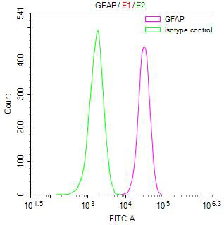
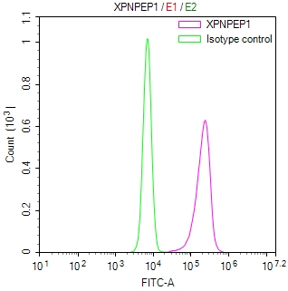
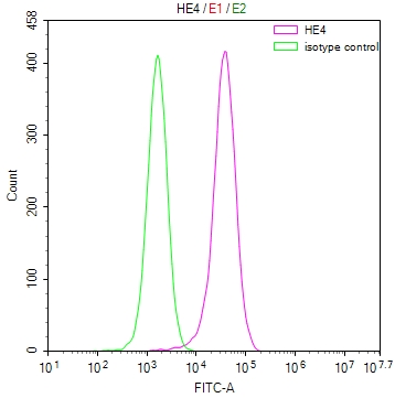
Overlay Peak curve showing Jurkat cells stained with CSB-RA009369MA1HU (red line) at 1:50. Then 10% normal goat serum was Incubated to block non-specific protein-protein interactions followed by the antibody (1µg/1*106cells) for 45 min at 4°C. The secondary antibody used was FITC-conjugated Goat Anti-Mouse IgG(H+L) at 1/200 dilution for 35 min at 4°C. Isotype control antibody (green line) was mouse IgG1 (1µg/1*106cells) used under the same conditions. Acquisition of >10,000 events was performed.
-
-
其他:
产品详情
-
Uniprot No.:P14136
-
基因名:
-
别名:GFAP antibody; GFAP Epsilon antibody; GFAP_HUMAN antibody; GFAPdelta antibody; GFAPepsilon antibody; Glial fibrillary acidic protein antibody; Intermediate filament protein antibody
-
反应种属:Human, Mouse, Rat
-
免疫原:Recombinant Human GFAP protein
-
免疫原种属:Homo sapiens (Human)
-
标记方式:Non-conjugated
-
克隆类型:Monoclonal
-
抗体亚型:Mouse IgG2a
-
纯化方式:Affinity-chromatography
-
克隆号:6B12
-
浓度:It differs from different batches. Please contact us to confirm it.
-
保存缓冲液:Preservative: 0.03% Proclin 300
Constituents: 50% Glycerol, 0.01M PBS, PH 7.4 -
产品提供形式:Liquid
-
应用范围:ELISA, WB, IHC, IF, FC
-
推荐稀释比:
Application Recommended Dilution WB 1:1000-1:5000 IHC 1:20-1:200 IF 1:20-1:200 FC 1:20-1:200 -
Protocols:
-
储存条件:Upon receipt, store at -20°C or -80°C. Avoid repeated freeze.
-
货期:Basically, we can dispatch the products out in 1-3 working days after receiving your orders. Delivery time maybe differs from different purchasing way or location, please kindly consult your local distributors for specific delivery time.
相关产品
靶点详情
-
功能:GFAP, a class-III intermediate filament, is a cell-specific marker that, during the development of the central nervous system, distinguishes astrocytes from other glial cells.
-
基因功能参考文献:
- The features of the neuropathology and immunopathology of GFAP astrocytopathies were perivascular inflammation and loss of astrocytes and neurons PMID: 29193473
- amniotic fluid -GFAP levels differentiate between myelomeningocele and myeloschisis, and raise interesting questions regarding the clinical significance between the 2 types of defects. PMID: 28768252
- Desmin, Glial Fibrillary Acidic Protein, Vimentin, and Peripherin are type III intermediate filaments that have roles in health and disease [review] PMID: 29196434
- Plasma concentration of GFAP demonstrated associations with stroke occurrence in a West African cohort but was not associated with stroke severity or mortality. PMID: 29074065
- This study demonstrated that Concentrations of microparticles expressing GFAP and AQP4 were significantly higher in the traumatic brain injury group compared with healthy controls. PMID: 28972406
- The s observed higher serum levels of GFAP and UCH-L1 in brain-injured children compared with controls and also demonstrated a step-wise increase of biomarker concentrations over the continuum of severity from mild to severe traumatic brain injury. Serum UCH-L1 and GFAP concentrations also strongly predicted poor outcome. PMID: 27319802
- Study examined if QKI6B expression can predict the outcome of GFAP, and several oligodendrocyte-related genes, in the prefrontal cortex of brain samples of schizophrenic individuals. QKI6B significantly predicts the expression of GFAP, but does not predict oligodendrocyte-related gene outcome, as previously seen with other QKI isoforms. PMID: 28552414
- GFAP, along with tau and AmyloidBeta42, were increased in plasma up to 90 days after traumatic brain injury compared with controls. PMID: 27312416
- Results show that the positive rates and expression levels of nestin, tyrosine hydroxylase (TH), GFAP and IL-17 were significantly decreased while Foxp3 and the ratio of Foxp3/IL-17 were statistically elevated in BM of AML patients. PMID: 27016413
- GFAP levels >0.29 ng/ml were seen only in intracerebral hemorrhage, thus confirming the diagnosis of ICH during prehospital care. PMID: 27951536
- These results indicate that autoantibodies against GFAP could serve as a predictive marker for the development of overt autoimmune diabetes. PMID: 28546444
- Higher median plasma GFAP values were documented in intracerebral hemorrhage compared with acute ischemic stroke, stroke mimics, and controls. PMID: 28751552
- GFAP is specifically expressed in the auricular chondrocytes, and assumes a pivotal role in resistance against mechanical stress. PMID: 28063220
- Bevacizumab treatment was also associated with structural protein abnormalities, with decreased GFAP and vimentin content and upregulated GFAP and vimentin mRNA expression. PMID: 28419863
- the exchange of GFP-GFAPdelta was significantly slower than the exchange of GFP-GFAPalpha with the intermediate filament-network. PMID: 27141937
- Tat expression or GFAP expression led to formation of GFAP aggregates and induction of unfolded protein response (UPR) and endoplasmic reticulum (ER) stress in astrocytes. PMID: 27609520
- This study demonstrated that GFAP exhibited distinct temporal profiles over the course of 7 days in patient with traumatic brain injury. PMID: 27018834
- e data indicates that serum GFAP levels may be associated with severity of autism spectrum disorders among Chinese children. PMID: 28088366
- High GFAP expression is associated with retinoblastoma. PMID: 27488116
- Overall, glial fibrillary acidic protein reflected no evidence for significant peripartum brain injury in neonates with congenital heart defects, but there was a trend for elevation by postnatal day 4 in neonates with left heart obstruction. PMID: 26786018
- serum levels of GFAP were significantly lower in autism spectrum disorders than controls PMID: 27097671
- We found downregulation of GFAP mRNA and protein in the mediodorsal thalamus and caudate nucleus of depressed suicides compared with controls, whereas GFAP expression in other brain regions was similar between groups. Furthermore, a regional comparison including all samples revealed that GFAP expression in both subcortical regions was, on average, between 11- and 15-fold greater than in cerebellum and neocortex. PMID: 26033239
- No difference in cord blood concentration found between hypoxic-ischemic encephalopathy neonates and controls PMID: 26135781
- GFAP is upregulated following an insult or injury to the brain, additionally making it an indicator of CNS pathology. PMID: 25846779
- This study demonistrated that the density of GFAP-immunoreactive astrocytes is decreased in left hippocampi in major depressive disorder PMID: 26742791
- This study demonstrated that GFAP as a promising biomarker to distinguish ischemic stroke from intracerebral hemorrhage. PMID: 26526443
- The levels of GFAP in Alzheimer's disease, dementia with Lewy bodies, and frontotemporal lobar degeneration patients were significantly higher than those in the healthy control subjects. PMID: 26485083
- GFAP is significantly associated with outcome, but it does not add predictive power to commonly used prognostic variables in a population of patients with TBI of varying severities. PMID: 26547005
- Neither duplications nor deletions of GFAP were found, suggesting that GFAP coding-region rearrangements may not be involved in Alexander disease or Alexanderrelated leukoencephalopathies. PMID: 26208460
- The data suggest that human vitreous body GFAP is a protein biomarker for glial activation in response to retinal pathologies. PMID: 26279003
- Studied diagnostic Value of Serum Levels of GFAP, pNF-H, and NSE Compared With Clinical Findings in Severity Assessment of Human Traumatic Spinal Cord Injury. PMID: 25341992
- GFAP peaks early during haemorrhagic brain lesions (at significantly higher levels), and late in ischaemic events, whereas antibodies against NR2 RNMDA have significantly higher levels during ischemic stroke at all time-points. PMID: 26081945
- There was an absence of GFAP in astrocytes during early fetal spinal cord development until 9 months of gestation , and the appearance of GFAP-positive reactivity was later than that of neurons. PMID: 25904356
- It could be a clinically relevant marker associated with tumor invasiveness in cerebral astrocytomas. PMID: 25178519
- These data imply that a tight regulation of histone acetylation in astrocytes is essential, because dysregulation of gene expression causes the aggregation of GFAP, a hallmark of human diseases like Alexander's disease. PMID: 25128567
- Identification of a novel nonsense mutation in the rod domain of GFAP that is associated with Alexander disease. PMID: 24755947
- The role of S100B protein, neuron-specific enolase, and glial fibrillary acidic protein in the evaluation of hypoxic brain injury in acute carbon monoxide poisoning PMID: 24505052
- GFAP, the principal intermediate filament protein of astrocytes, is involved in physiological, but in particular, in pathophysiological functions of astrocytes, the latter ones being connected with astrocyte activation and reactive gliosis. [Review] PMID: 25726916
- The data on the changes in expression of GFAP in Alexander disease caused by the primary pathology of astrocytes are presented. PMID: 25859599
- A combined profile of preoperative IGFBP-2, GFAP, and YKL-40 plasma levels could serve as an additional diagnostic tool for patients with inoperable brain lesions suggestive of Glioblastoma multiforme. PMID: 25139333
- There are significant increases in glial fibrillary acidic protein levels in children undergoing cardiopulmonary bypass for repair of congenital heart disease. The highest values were seen during the re-warming phase. PMID: 23845562
- This stuidy demonistrated that Fibrillary astrocytes are decreased in the subgenual cingulate in schizophrenia. PMID: 24374936
- TBI patients showed an average 3.77 fold increase in anti-GFAP autoantibody levels from early (0-1 days) to late (7-10 days) times post injury. PMID: 24667434
- We showed that GFAP is over-expressed and hypophosphorylated in the enteric glial cells of Parkinson's disease patients as compared to healthy subjects PMID: 24749759
- Its expression is associated with plaque load related astrogliosis in Alzheimer's disease. PMID: 24269023
- The findings of this study that caspase-mediated GFAP proteolysis may be a common event in the context of both the GFAP mutation and excess. PMID: 24102621
- This study demonistratedt hat Increased expression of glial fibrillary acidic protein in prefrontal cortex in psychotic illness PMID: 23911257
- Data indicate that Gfapdelta is expressed in the in developing mouse brain sub-ventricular zones in accordance with the described localization in the developing and adult human brain. PMID: 23991052
- GFAP-breakdown products blood levels reliably distinguished severity of injury in traumatic brain injury patients. PMID: 23489259
- The C/C genotype at rs2070935 of the GFAP promoter in late-onset AxD was associated with an earlier onset and a more rapid progression of ambulatory disability compared with the other genotypes. PMID: 23903069
显示更多
收起更多
-
相关疾病:Alexander disease (ALXDRD)
-
亚细胞定位:Cytoplasm.
-
蛋白家族:Intermediate filament family
-
组织特异性:Expressed in cells lacking fibronectin.
-
数据库链接:
HGNC: 4235
OMIM: 137780
KEGG: hsa:2670
STRING: 9606.ENSP00000253408
UniGene: Hs.514227
Most popular with customers
-
-
YWHAB Recombinant Monoclonal Antibody
Applications: ELISA, WB, IF, FC
Species Reactivity: Human, Mouse, Rat
-
Phospho-YAP1 (S127) Recombinant Monoclonal Antibody
Applications: ELISA, WB, IHC
Species Reactivity: Human
-
-
-
-
-


