UCP1 Antibody
-
货号:CSB-PA599294GA01HU
-
规格:¥3,900
-
图片:
-
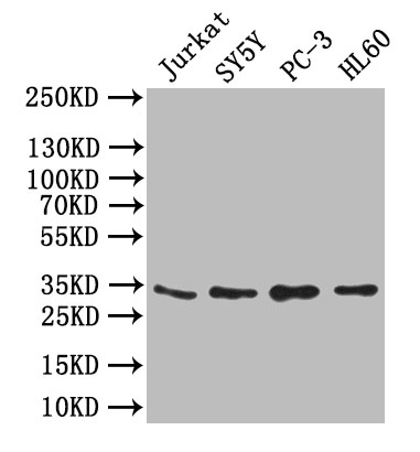
Western Blot
Positive WB detected in: JK whole cell lysate,SY5Y whole cell lysate,PC-3 whole cell lysate,,HL60 whole cell lysate
All lanes: UCP1 antibody at 1:1000
Secondary
Goat polyclonal to rabbit IgG at 1/50000 dilution
Predicted band size: 33 kDa
Observed band size: 33 kDa -
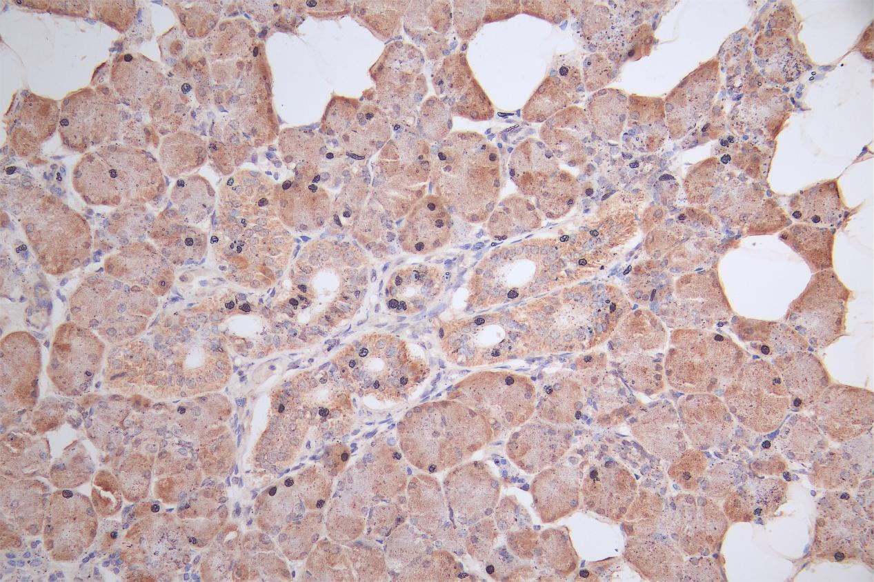
IHC image of CSB-PA025554LA01HU diluted at 1:50 and staining in paraffin-embedded human salivary gland tissue performed on a Leica BondTM system. After dewaxing and hydration, antigen retrieval was mediated by high pressure in a citrate buffer (pH 6.0). Section was blocked with 10% normal goat serum 30min at RT. Then primary antibody (1% BSA) was incubated at 4°C overnight. The primary is detected by a Goat anti-rabbit polymer IgG labeled by HRP and visualized using 0.05% DAB.
-
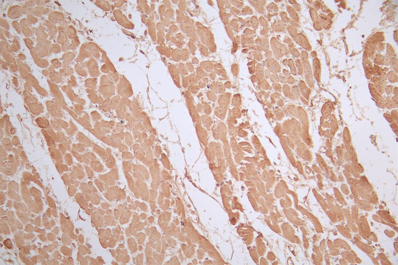
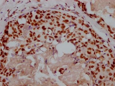
IHC image of CSB-PA025554LA01HU diluted at 1:50 and staining in paraffin-embedded human heart tissue performed on a Leica BondTM system. After dewaxing and hydration, antigen retrieval was mediated by high pressure in a citrate buffer (pH 6.0). Section was blocked with 10% normal goat serum 30min at RT. Then primary antibody (1% BSA) was incubated at 4°C overnight. The primary is detected by a Goat anti-rabbit polymer IgG labeled by HRP and visualized using 0.05% DAB.
-
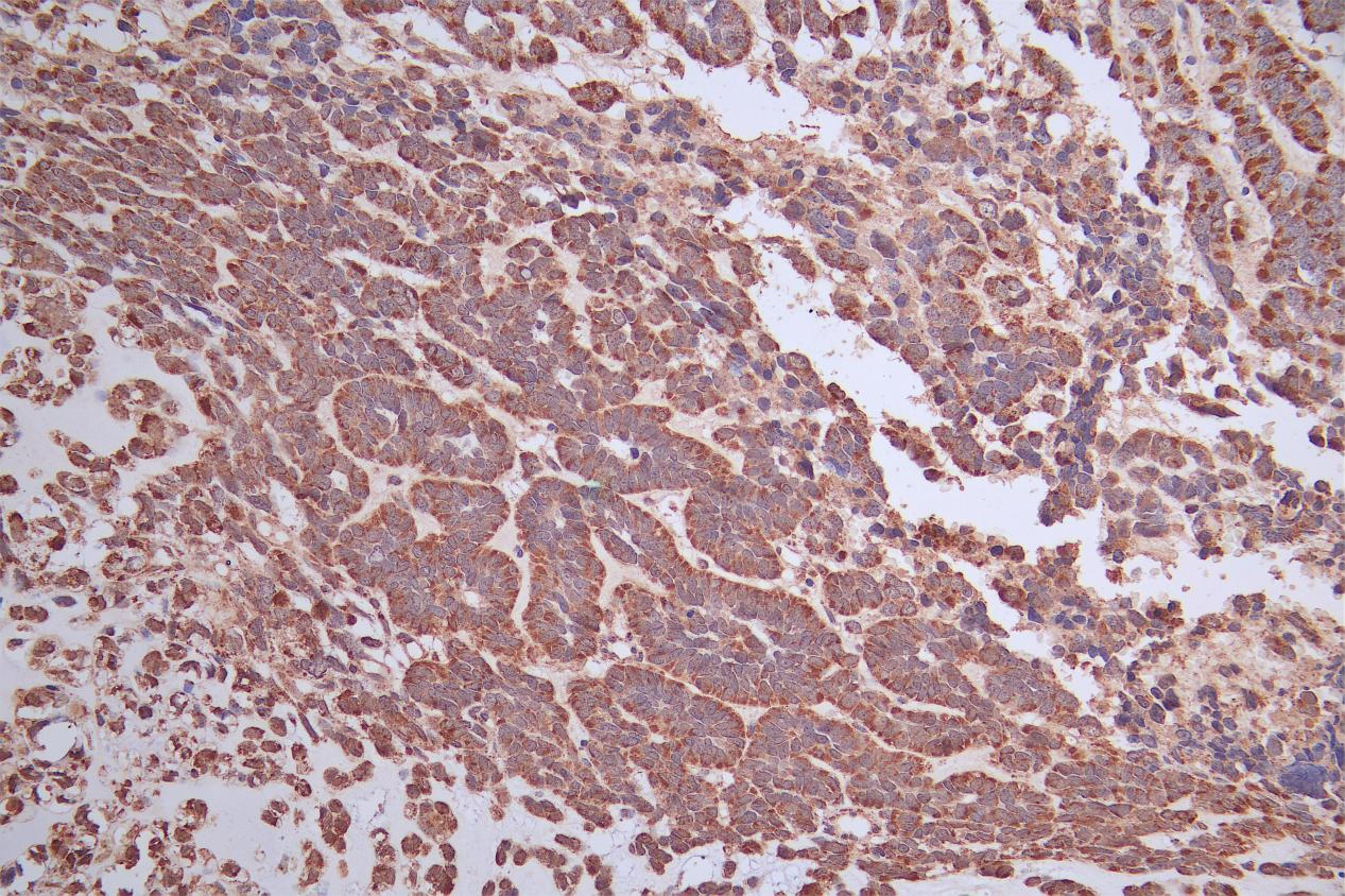
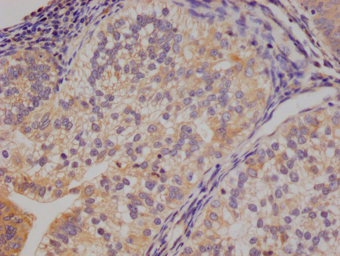
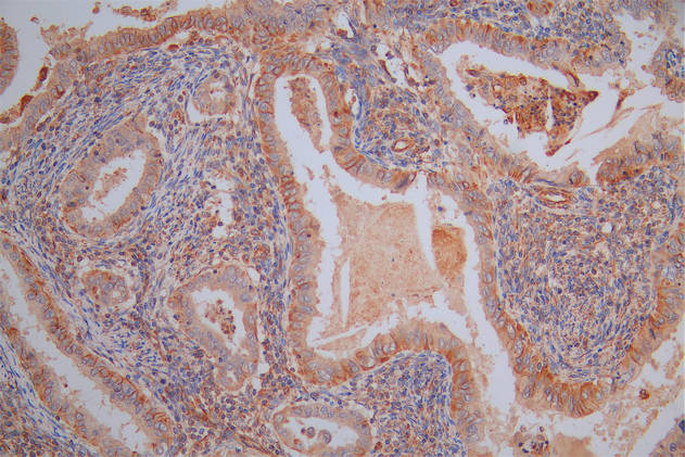
IHC image of CSB-PA025554LA01HU diluted at 1:50 and staining in paraffin-embedded human ovarian cancer performed on a Leica BondTM system. After dewaxing and hydration, antigen retrieval was mediated by high pressure in a citrate buffer (pH 6.0). Section was blocked with 10% normal goat serum 30min at RT. Then primary antibody (1% BSA) was incubated at 4°C overnight. The primary is detected by a Goat anti-rabbit polymer IgG labeled by HRP and visualized using 0.05% DAB.
-
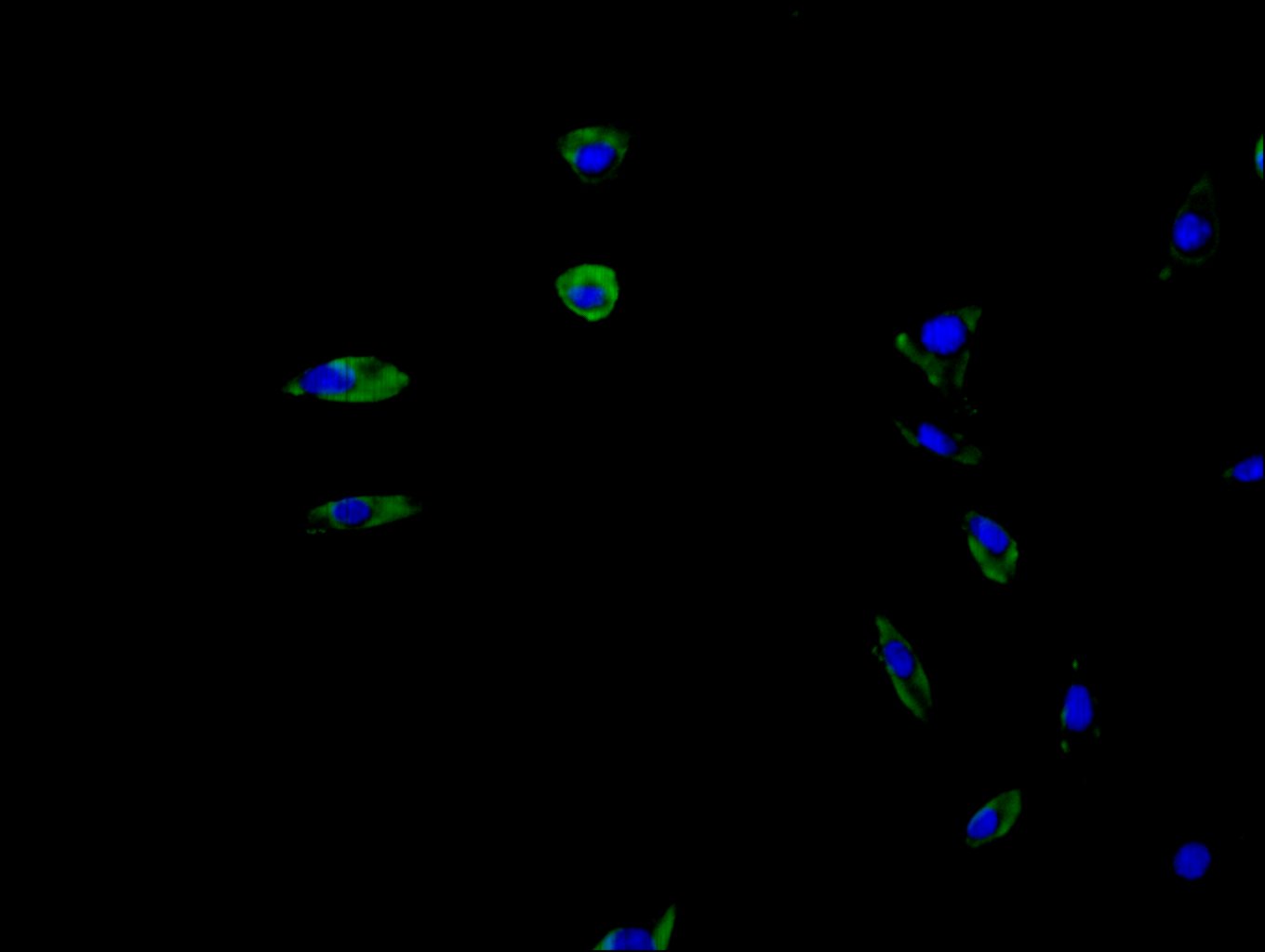
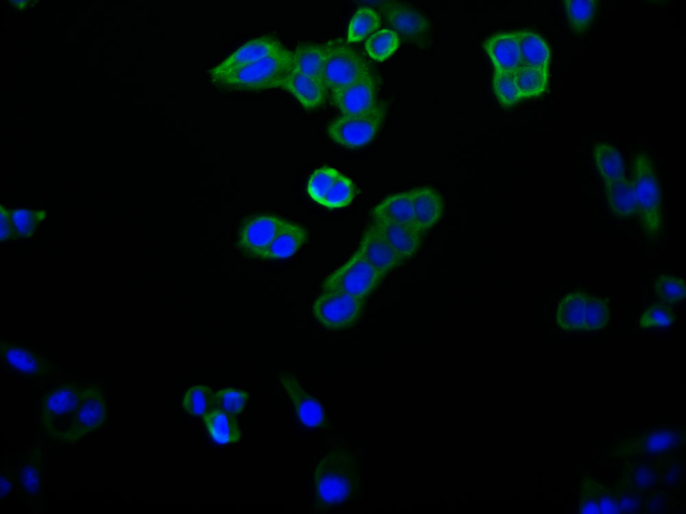
Immunofluorescence staining of SH-SY5Y cell with CSB-PA025554LA01HU at 1:20, counter-stained with DAPI. The cells were fixed in 4% formaldehyde and blocked in 10% normal Goat Serum. The cells were then incubated with the antibody overnight at 4C. The secondary antibody was Alexa Fluor 488-congugated AffiniPure Goat Anti-Rabbit IgG(H+L).
-
-
其他:
产品详情
-
产品名称:Rabbit anti-Homo sapiens (Human) UCP1 Polyclonal antibody
-
Uniprot No.:Q9BRC7
-
基因名:
-
别名:mitochondrial brown fat uncoupling protein antibody; Mitochondrial brown fat uncoupling protein 1 antibody; SLC25A7 antibody; Solute carrier family 25 member 7 antibody; Thermogenin antibody; UCP 1 antibody; UCP antibody; UCP1 antibody; UCP1_HUMAN antibody; uncoupling protein 1 (mitochondrial, proton carrier) antibody; Uncoupling protein 1 antibody
-
宿主:Rabbit
-
反应种属:Human,Mouse,Rat
-
免疫原:Recombinant Human Mitochondrial brown fat uncoupling protein 1 protein (1-307AA)
-
免疫原种属:Homo sapiens (Human)
-
标记方式:Non-conjugated
-
克隆类型:Polyclonal
-
抗体亚型:IgG
-
纯化方式:Antigen Affinity Purified
-
浓度:It differs from different batches. Please contact us to confirm it.
-
保存缓冲液:PBS with 0.1% Sodium Azide, 50% Glycerol, pH 7.3. -20°C, Avoid freeze / thaw cycles.
-
产品提供形式:Liquid
-
应用范围:ELISA, WB, IHC, IF
-
推荐稀释比:
Application Recommended Dilution WB 1:1000-1:5000 IHC 1:50-1:500 IF 1:20-1:200 -
Protocols:
-
储存条件:Upon receipt, store at -20°C or -80°C. Avoid repeated freeze.
-
货期:Basically, we can dispatch the products out in 1-3 working days after receiving your orders. Delivery time maybe differs from different purchasing way or location, please kindly consult your local distributors for specific delivery time.
相关产品
靶点详情
-
功能:Hydrolyzes the phosphatidylinositol 4,5-bisphosphate (PIP2) to generate 2 second messenger molecules diacylglycerol (DAG) and inositol 1,4,5-trisphosphate (IP3). DAG mediates the activation of protein kinase C (PKC), while IP3 releases Ca(2+) from intracellular stores. Required for acrosome reaction in sperm during fertilization, probably by acting as an important enzyme for intracellular Ca(2+) mobilization in the zona pellucida-induced acrosome reaction. May play a role in cell growth. Modulates the liver regeneration in cooperation with nuclear PKC. Overexpression up-regulates the Erk signaling pathway and proliferation.; Acts as a non-receptor guanine nucleotide exchange factor which binds to and activates guanine nucleotide-binding protein (G-protein) alpha subunit GNAI3.
-
基因功能参考文献:
- Here, the s report that the zebrafish/human phosphatidylinositol transfer protein Sec14l3/SEC14L2 act as GTPase proteins to transduce Wnt signals from Frizzled to phospholipase C (PLC). PMID: 28463110
- PLCdelta(1) and PLCdelta(4) are probably differentially regulated in distinct cellular compartments by PI(4,5)P(2) and the PH domain of PLCdelta(4) does not act as a localization signal PMID: 15037625
- Serial deletion analysis identified the core PLC-delta4 promoter region as being between -402 and -67, in which an E-box and an AP-1 binding site played important roles in the promoter activity. PMID: 17394098
-
亚细胞定位:Membrane; Peripheral membrane protein. Nucleus. Cytoplasm. Endoplasmic reticulum. Note=Localizes primarily to intracellular membranes mostly to the endoplasmic reticulum.
-
组织特异性:Highly expressed in skeletal muscle and kidney tissues, and at moderate level in intestinal tissue. Expressed in corneal epithelial cells.
-
数据库链接:
HGNC: 9062
OMIM: 605939
KEGG: hsa:84812
STRING: 9606.ENSP00000388631
UniGene: Hs.632528
Most popular with customers
-
-
YWHAB Recombinant Monoclonal Antibody
Applications: ELISA, WB, IF, FC
Species Reactivity: Human, Mouse, Rat
-
-
-
-
-
-

