CD63 Monoclonal Antibody
-
货号:CSB-MA004950A0m
-
规格:¥1320
-
图片:
-
Western Blot
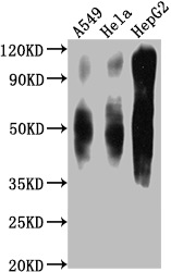
Positive WB detected in: A549 whole cell lysate, Hela whole cell lysate, HepG2 whole cell lysate
All lanes CD63 antibody at 1:1000
Secondary
Goat polyclonal to mouse IgG at 1/50000 dilution
Predicted band size: 30-120 KD KDa
Observed band size: 30-120 KD KDa
Exposure time:1min -
Western Blot
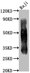
Positive WB detected in: Raji whole cell lysate
All lanes CD63 antibody at 1:1000
Secondary
Goat polyclonal to mouse IgG at 1/50000 dilution
Predicted band size: 30-120 KD KDa
Observed band size: 30-120 KD KDa
Exposure time:1min -
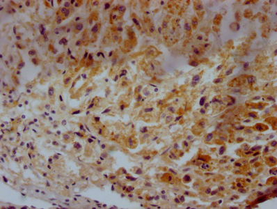
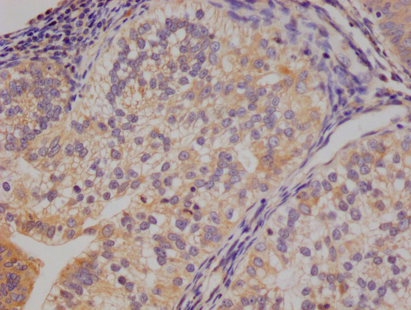
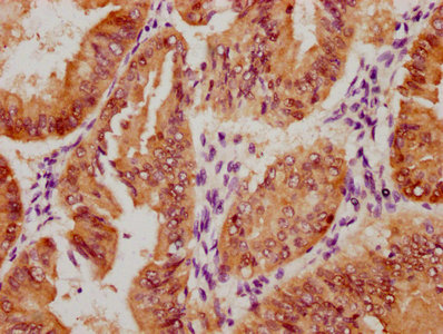
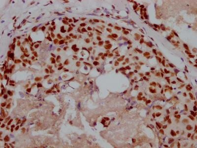
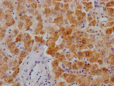
IHC image of CSB-MA004950A0m diluted at 1:500 and staining in paraffin-embedded human glioma tissue performed on a Leica BondTM system. After dewaxing and hydration, antigen retrieval was mediated by high pressure in a citrate buffer (pH 6.0). Section was blocked with 10% normal goat serum 30min at 37°C. Then primary antibody (1% BSA) was incubated at 4°C overnight. The primary is detected by a Goat anti-rabbit IgG labeled by HRP and visualized using 0.05% DAB.
-
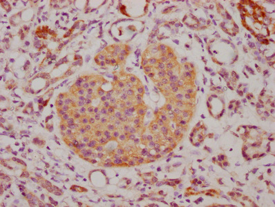
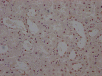
IHC image of CSB-MA004950A0m diluted at 1:500 and staining in paraffin-embedded human lung cancer tissue performed on a Leica BondTM system. After dewaxing and hydration, antigen retrieval was mediated by high pressure in a citrate buffer (pH 6.0). Section was blocked with 10% normal goat serum 30min at 37°C. Then primary antibody (1% BSA) was incubated at 4°C overnight. The primary is detected by a Goat anti-rabbit IgG labeled by HRP and visualized using 0.05% DAB.
-
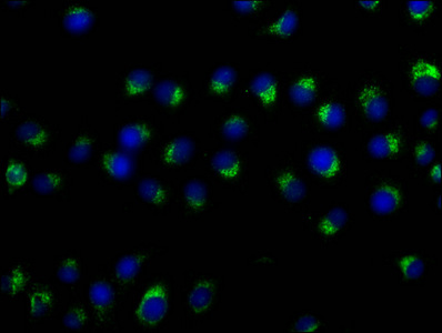
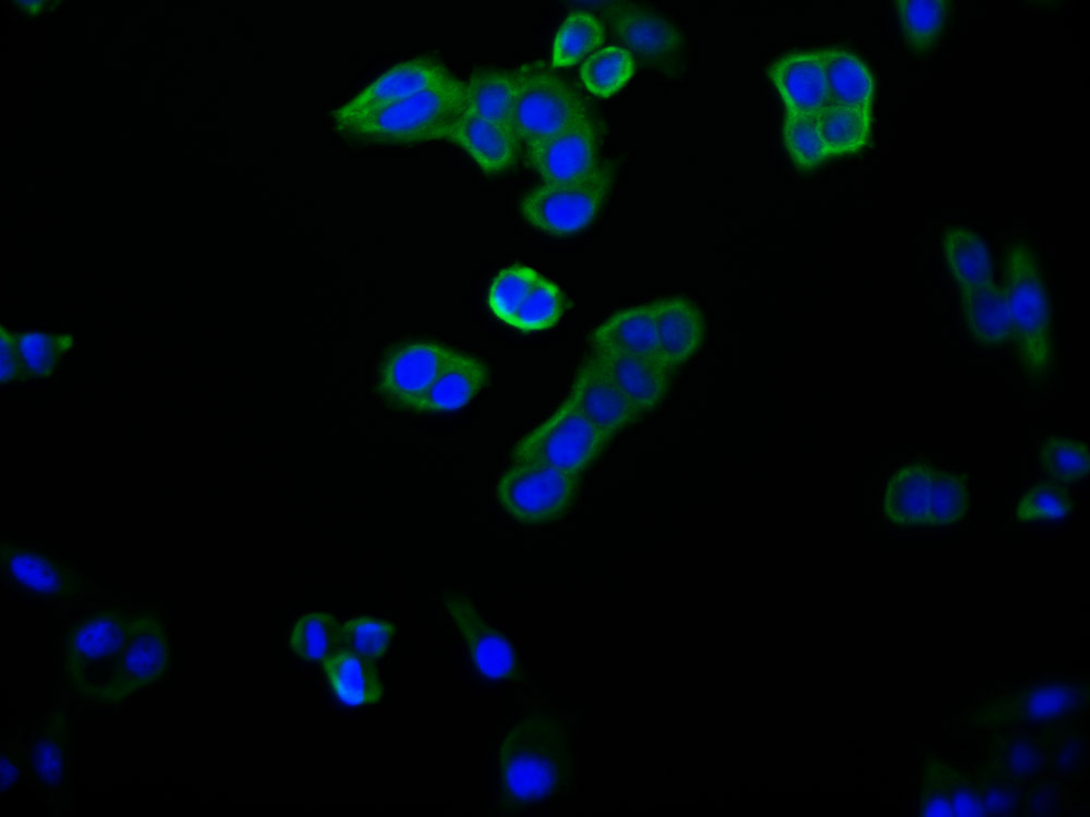
Immunofluorescence staining of A549 cells with CSB-MA004950A0m at 1:50, counter-stained with DAPI. The cells were fixed in 4% formaldehyde and blocked in 10% normal Goat Serum. The cells were then incubated with the antibody overnight at 4°C. Nuclear DNA was labeled in blue with DAPI. The secondary antibody was FITC-conjugated AffiniPure Goat Anti-Mouse IgG (H+L).
-
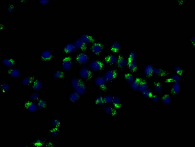
Immunofluorescence staining of Hela cells with CSB-MA004950A0m at 1:50, counter-stained with DAPI. The cells were fixed in 4% formaldehyde and blocked in 10% normal Goat Serum. The cells were then incubated with the antibody overnight at 4°C. Nuclear DNA was labeled in blue with DAPI. The secondary antibody was FITC-conjugated AffiniPure Goat Anti-Mouse IgG (H+L).
-
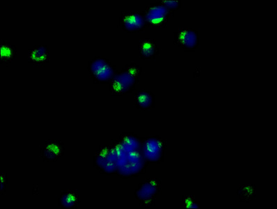
Immunofluorescence staining of MCF-7 cells with CSB-MA004950A0m at 1:50, counter-stained with DAPI. The cells were fixed in 4% formaldehyde and blocked in 10% normal Goat Serum. The cells were then incubated with the antibody overnight at 4°C. Nuclear DNA was labeled in blue with DAPI. The secondary antibody was FITC-conjugated AffiniPure Goat Anti-Mouse IgG (H+L).
-
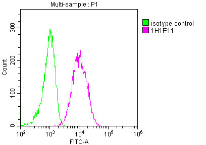
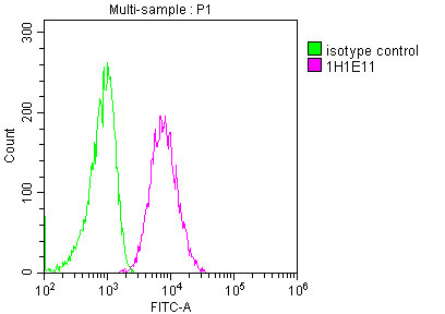
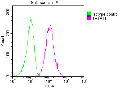
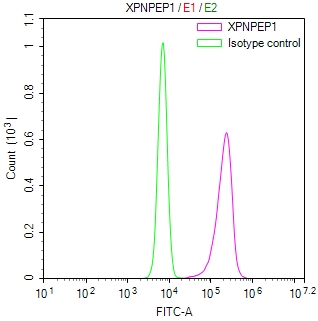
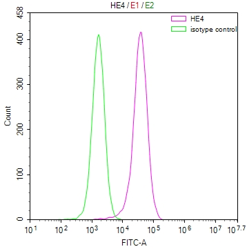
Overlay histogram showing A549 cells stained with CSB-MA004950A0m (red line) at 1:100. The cells were fixed in 4% formaldehyde and permeated by 0.2% TritonX-100. Then 10% normal goat serum was Incubated to block non-specific protein-protein interactions followed by the antibody (1µg/1*106cells) for 1 h at 4°C. The secondary antibody used was FITC-conjugated Goat Anti-Mouse IgG(H+L) at 1/100 dilution for 30min at 4°C. Isotype control antibody (green line) was mouse IgG2b (1µg/1*106cells) used under the same conditions. Acquisition of >10,000 events was performed.
-
Overlay histogram showing Hela cells stained with CSB-MA004950A0m (red line) at 1:100. The cells were fixed in 4% formaldehyde and permeated by 0.2% TritonX-100. Then 10% normal goat serum was Incubated to block non-specific protein-protein interactions followed by the antibody (1µg/1*106cells) for 1 h at 4°C. The secondary antibody used was FITC-conjugated Goat Anti-Mouse IgG(H+L) at 1/100 dilution for 30min at 4°C. Isotype control antibody (green line) was mouse IgG2b (1µg/1*106cells) used under the same conditions. Acquisition of >10,000 events was performed.
-
Overlay histogram showing HepG2 cells stained with CSB-MA004950A0m (red line) at 1:100. The cells were fixed in 4% formaldehyde and permeated by 0.2% TritonX-100. Then 10% normal goat serum was Incubated to block non-specific protein-protein interactions followed by the antibody (1µg/1*106cells) for 1 h at 4°C. The secondary antibody used was FITC-conjugated Goat Anti-Mouse IgG(H+L) at 1/100 dilution for 30min at 4°C. Isotype control antibody (green line) was mouse IgG2b (1µg/1*106cells) used under the same conditions. Acquisition of >10,000 events was performed.
-
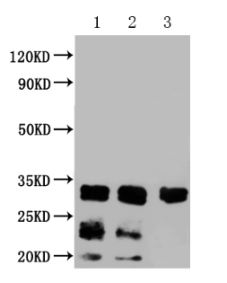
1. Exosomes extracted from Hela cells
2. Exosomes extracted from Hela cells
3. Exosomes extracted from urine
-
-
其他:
产品详情
-
产品描述:
CD63 monoclonal antibody is an IgG2a isotype that specifically recognizes CD63 protein. It has been extensively characterized and shown to be highly specific in human samples. This CD63 monoclonal antibody is suitable for use in a variety of research applications, including ELISA, WB, IHC, IF, and FC. It was generated using hybridoma technology by fusing mouse spleen cells immunized with recombinant human CD63 protein (103-203aa) with myeloma cells. The resulting hybridoma clones were screened for those that produce high-affinity CD63 antibodies. The selected hybridomas are cultured in the mouse abdominal cavity, and CD63 monoclonal antibodies are then collected from the mouse ascites.
CD63, also known as LAMP-3, is primarily found in the membranes of late endosomes and lysosomes and is involved in various cellular processes such as antigen presentation, phagocytosis, and intracellular trafficking. CD63 interacts with other tetraspanins, integrins, and other proteins to form tetraspanin-enriched microdomains (TEMs) in the membrane. These TEMs play a role in signal transduction, protein trafficking, and cell adhesion.
-
产品名称:Mouse anti-Homo sapiens (Human) CD63 Monoclonal Antibody antibody
-
Uniprot No.:P08962
-
基因名:CD63
-
别名:CD63; MLA1; TSPAN30; CD63 antigen; Granulophysin; Lysosomal-associated membrane protein 3; LAMP-3; Lysosome integral membrane protein 1; Limp1; Melanoma-associated antigen ME491; OMA81H; Ocular melanoma-associated antigen; Tetraspanin-30; Tspan-30; CD antigen CD63
-
宿主:Mouse
-
反应种属:Human
-
免疫原:Recombinant Human CD63 antigen protein (103-203AA)
-
免疫原种属:Homo sapiens (Human)
-
标记方式:Non-conjugated
-
克隆类型:Monoclonal Antibody
-
抗体亚型:IgG2a
-
纯化方式:>95%, Protein A purified
-
克隆号:1H1E11
-
浓度:It differs from different batches. Please contact us to confirm it.
-
保存缓冲液:Preservative: 0.03% Proclin 300
Constituents: 50% Glycerol, 0.01M PBS, PH 7.4 -
产品提供形式:Liquid
-
应用范围:ELISA, WB, IHC, IF, FC
-
推荐稀释比:
Application Recommended Dilution WB WB:1:1000-1:8000 IHC 1:50-1:200 IF 1:50-1:200 FC 1:50-1:200 -
Protocols:
-
储存条件:Upon receipt, store at -20°C or -80°C. Avoid repeated freeze.
-
货期:Basically, we can dispatch the products out in 1-3 working days after receiving your orders. Delivery time maybe differs from different purchasing way or location, please kindly consult your local distributors for specific delivery time.
相关产品
靶点详情
-
功能:Functions as cell surface receptor for TIMP1 and plays a role in the activation of cellular signaling cascades. Plays a role in the activation of ITGB1 and integrin signaling, leading to the activation of AKT, FAK/PTK2 and MAP kinases. Promotes cell survival, reorganization of the actin cytoskeleton, cell adhesion, spreading and migration, via its role in the activation of AKT and FAK/PTK2. Plays a role in VEGFA signaling via its role in regulating the internalization of KDR/VEGFR2. Plays a role in intracellular vesicular transport processes, and is required for normal trafficking of the PMEL luminal domain that is essential for the development and maturation of melanocytes. Plays a role in the adhesion of leukocytes onto endothelial cells via its role in the regulation of SELP trafficking. May play a role in mast cell degranulation in response to Ms4a2/FceRI stimulation, but not in mast cell degranulation in response to other stimuli.
-
基因功能参考文献:
- Signaling focal contacts via CD63 were identified to facilitate cell adhesion, migration and mediate extracellular matrix-physical cues to modulate hematopoietic stem cells and progenitor cells function. PMID: 28566689
- Human CD63-GFP expression was controlled under the rat Sox2 promoter (Sox2/human CD63-GFP), and it was expressed in undifferentiated fetal brains. PMID: 29208635
- our results identify the CD63-syntenin-1-ALIX complex as a key regulatory component in post-endocytic HPV trafficking. PMID: 27578500
- CD63 and exosome expression is altered in scleroderma dermal fibroblasts PMID: 27443953
- CD63 is a critical player in LMP1 exosomal trafficking and LMP1-mediated enhancement of exosome production and may play further roles in limiting downstream LMP1 signaling. PMID: 27974566
- TIMP1 signaling via CD63 leads to activation of hepatic stellate cells, which create an environment in the liver that increases its susceptibility to pancreatic tumor cells. PMID: 27506299
- Concentrations of Cd63 were elevated in gingival crevicular fluid of patients with pre-eclampsia. PMID: 26988336
- CD163(+) TAMs in oral premalignant lesions coexpress CD163 and STAT1, suggesting that the TAMs in oral premalignant lesions possess an M1 phenotype in a Th1-dominated micromilieu. PMID: 26242181
- Our findings reveal that elevated levels of TIMP-1 impact on neutrophil homeostasis via signaling through CD63. PMID: 26001794
- These findings indicate that rhTIMP-1 promotes clonogenic expansion and survival in human progenitors via the activation of the CD63/PI3K/pAkt signaling pathway PMID: 26213230
- It was shown that treatment of macrophages with anti-CD63 inhibits CCR5-mediated virus infection in a cell type-specific manner, but that no inhibition of CXCR4-tropic viruses occurs. PMID: 25658293
- TM4SF5 interacted with CD151, and caused the internalization of CD63 from the cell surface. PMID: 25033048
- Diesel exhaust exposure during exercise induces platelet activation as illustrated by a dose-response increase in the release of CD62P and CD63. PMID: 25297946
- Data show that CD63 is a crucial player in the regulation of the tumor cell-intrinsic metastatic potential by affecting cell plasticity. PMID: 25354204
- CD63 tetraspanin is a negative driver of epithelial-to-mesenchymal transition in human melanoma cells. PMID: 24940653
- these results indicated that high glycosylation of CD63 by RPN2 is implicated in clinical outcomes in breast cancer patients PMID: 24884960
- Collectively, these findings indicate that CD63 may support Env-mediated fusion as well as a late (post-integration) step in the HIV-1 replication cycle. PMID: 24507450
- Data suggest that TIMP1 (tissue inhibitor of metalloproteinase-1) acts as chemoattractant for neural stem cells (NSC); TIMP1 enhances NSC adhesion, migration, and cytoskeletal reorganization; these effects are dependent on CD63/CD29 (integrin beta1). PMID: 24635319
- Timp1 is assembled in a supramolecular complex containing CD63 and beta1-integrins along melanoma genesis and confers anoikis resistance by activating PI3-K signaling pathway. PMID: 23522389
- Antigen-induced p38 MAPK phosphorylation in human basophils essentially contributes to CD63 upregulation PMID: 18727065
- Loss of CD63 has a similar phenotype to loss of P-selectin itself, thus CD63 is an essential cofactor to P-selectin. PMID: 21803846
- shRNA knockdown in B lymphoblastoid cell line results to increased CD4(+) T-cell recognition PMID: 21660937
- decreased levels of CD63 were associated with distant and lymph node metastasis status and does play a direct role in human gastric carcinogenesis. PMID: 21521534
- C-terminal modifications that retain LMP1 in Golgi compartments preclude assembly within CD63-enriched domains and/or exosomal discharge leading to NF-kappaB overstimulation. PMID: 21527913
- These findings suggest that CD63 plays an early post-entry role prior to or at the reverse transcription step. PMID: 21315401
- ameloblastin is expressed in osteoblasts and functions as a promoting factor for osteogenic differentiation via a novel pathway through the interaction between CD63 and integrin beta1 PMID: 21149578
- Surface expression of the novel CD63 variant is a distinguishing feature of mast cells, which are are stable, multiple-use cells capable of surviving and delivering several consecutive hits PMID: 20337613
- this work provides the first evidence of a TIMP-4/CD63 association in astrocytoma tumor cells PMID: 20693981
- CD63 expression results from only the anaphylactic degranulation form of histamine release. PMID: 20633031
- Serum sCD163 is a homogenous protein covering more than 94% of the CD163 ectodomain including the haptoglobin-hemoglobin -binding region. PMID: 19581020
- Results show that AP-3 is absolutely required for the delivery of CD63 to lysosomes via the trans-Golgi network. PMID: 11907283
- downregulation of CD63 antigen is associated with breast tumor progression PMID: 12579280
- possible role in HIV-1 infections specific for macrophages PMID: 12610138
- Post-translational modification of CD63 may be involved in the functional and morphological changes of MHC class II compartments that occur during dendritic cell maturation PMID: 12755696
- relationships between the expression levels of CD61, CD63, and PAC-1 on the platelet surface and the incidences of acute rejection and tubular necrosis as well as the recovery of graft function after renal transplantation PMID: 12826159
- Upon platelet interaction with fibrinogen, cholesterol accumulated at the tips of filopodia and at the leading edge of spreading cells; cholesterol-rich raft aggregation was accompanied by concentration of c-Src and CD63 in these cell domains PMID: 12871315
- The study on CD63 included its chemistry eg. if it had and O-linked carbohydrate that was digested with O-glycanase. PMID: 12974720
- CD63 serves as an adaptor protein that links its interaction partners to the endocytic machinery of the cell. PMID: 14660791
- results suggest that CD9, CD63, CD81, and CD82 could play a role in modulating the interactions between immature DCs and their environment, slowing their migratory ability. However, only CD63 would intervene in the internalization of complex antigens PMID: 15130945
- Constitutive expression of CD63 may indicate that this factor does not play a direct role in thyroid carcinogenesis PMID: 15375577
- CD63 represents an activation-induced reinforcing element, whose triggering promotes sustained and efficient T cell activation and expansion. PMID: 15528334
- The linkage of CD63 with PI 4-kinase may result in the recruitment of this signaling enzyme to specific membrane locations in the platelet where it influences phosphoinositide-dependent signaling and platelet spreading. PMID: 15711748
- This study identifies a trafficking pathway from CD63-positive multivesicular bodies to the bacterial inclusion, a novel interaction that provides essential lipids necessary for maintenance of a productive intracellular infection. PMID: 16410552
- CD63 is recruited to already-budded Weibel-Palade bodies by an AP-3-dependent route PMID: 16683915
- CD63-syntenin-1 complex is abundant on the plasma membrane PMID: 16908530
- CD63 is a cell surface binding partner for TIMP-1, regulating cell survival and polarization via TIMP-1 modulation of tetraspanin/integrin signaling complex. PMID: 16917503
- Chronic urticaria serum-induced CD63 expression assay performed on atopic whole blood by means of tricolor flow cytometry could be the most useful tool for identification of a subset of patients with autoimmune chronic urticaria. PMID: 16918509
- In conclusion, using well-defined experimental conditions, the measurement of CD203c up-regulation on basophils in response to specific allergens is as reliable as CD63-BAT for the in vitro diagnosis of patients with IgE-mediated allergy. PMID: 17275019
- positive correlation between CD63 and erythrocyte sedimentation rate in rheumatoid arthritis PMID: 17279322
- Results suggest that CD63 can be a biomarker for predicting the prognosis in early stages of lung adenocarcinoma. PMID: 17350713
显示更多
收起更多
-
亚细胞定位:Cell membrane; Multi-pass membrane protein. Lysosome membrane; Multi-pass membrane protein. Late endosome membrane; Multi-pass membrane protein. Endosome, multivesicular body. Melanosome. Secreted, extracellular exosome. Cell surface.
-
蛋白家族:Tetraspanin (TM4SF) family
-
组织特异性:Detected in platelets (at protein level). Dysplastic nevi, radial growth phase primary melanomas, hematopoietic cells, tissue macrophages.
-
数据库链接:
HGNC: 1692
OMIM: 155740
KEGG: hsa:967
STRING: 9606.ENSP00000257857
UniGene: Hs.445570
Most popular with customers
-
-
YWHAB Recombinant Monoclonal Antibody
Applications: ELISA, WB, IF, FC
Species Reactivity: Human, Mouse, Rat
-
Phospho-YAP1 (S127) Recombinant Monoclonal Antibody
Applications: ELISA, WB, IHC
Species Reactivity: Human
-
-
-
-
-

