-
中文名称:小鼠钙黏蛋白5(CDH5)酶联免疫试剂盒
-
货号:CSB-EL005054MO
-
规格:96T/48T
-
价格:¥3600/¥2500
-
其他:
产品详情
-
产品描述:
This Mouse CDH5 ELISA Kit was designed for the quantitative measurement of Mouse CDH5 protein in serum, plasma, tissue homogenates. It is a Sandwich ELISA kit, its detection range is 0.235 ng/mL-15 ng/mL and the sensitivity is 0.058 ng/mL.
-
别名:Cdh5; Cadherin-5; Vascular endothelial cadherin; VE-cadherin; CD antigen CD144
-
缩写:
-
Uniprot No.:
-
种属:Mus musculus (Mouse)
-
样本类型:serum, plasma, tissue homogenates
-
检测范围:0.235 ng/mL-15 ng/mL
-
灵敏度:0.058 ng/mL
-
反应时间:1-5h
-
样本体积:50-100ul
-
检测波长:450 nm
-
研究领域:Cancer
-
测定原理:quantitative
-
测定方法:Sandwich
-
精密度:
Intra-assay Precision (Precision within an assay): CV%<8% Three samples of known concentration were tested twenty times on one plate to assess. Inter-assay Precision (Precision between assays): CV%<10% Three samples of known concentration were tested in twenty assays to assess. -
线性度:
To assess the linearity of the assay, samples were spiked with high concentrations of mouse CDH5 in various matrices and diluted with the Sample Diluent to produce samples with values within the dynamic range of the assay. Sample Serum(n=4) 1:100 Average % 92 Range % 88-96 1:200 Average % 95 Range % 91-98 1:400 Average % 100 Range % 96-105 1:800 Average % 94 Range % 87-98 -
回收率:
The recovery of mouse CDH5 spiked to levels throughout the range of the assay in various matrices was evaluated. Samples were diluted prior to assay as directed in the Sample Preparation section. Sample Type Average % Recovery Range Serum (n=5) 99 92-108 EDTA plasma (n=4) 94 90-98 -
标准曲线:
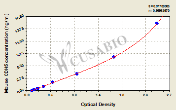
These standard curves are provided for demonstration only. A standard curve should be generated for each set of samples assayed. 
ng/ml OD1 OD2 Average Corrected 15 2.513 2.413 2.463 2.359 7.5 1.698 1.598 1.648 1.544 3.75 0.970 0.940 0.955 0.851 1.88 0.499 0.489 0.494 0.390 0.94 0.326 0.316 0.321 0.217 0.47 0.217 0.207 0.212 0.108 0.235 0.147 0.145 0.146 0.042 0 0.105 0.103 0.104 -
数据处理:
-
货期:3-5 working days
引用文献
- Application of multifunctional targeting epirubicin liposomes in the treatment of non-small-cell lung cancer. Song XL.et al,Int J Nanomedicine.,2017
- Nonhematopoietic Nrf2 dominantly impedes adult progression of sickle cell anemia in mice. Ghosh S.et al,JCI Insight.,2016
- The Antiatherogenic Effect of Fish Oil in Male Mice Is Associated with a Diminished Release of Endothelial ADAM17 and ADAM10 Substrates Speck N.et al,J Nutr.,2015
相关产品
靶点详情
-
功能:Cadherins are calcium-dependent cell adhesion proteins. They preferentially interact with themselves in a homophilic manner in connecting cells; cadherins may thus contribute to the sorting of heterogeneous cell types. This cadherin may play an important role in endothelial cell biology through control of the cohesion and organization of the intercellular junctions. It associates with alpha-catenin forming a link to the cytoskeleton. Acts in concert with KRIT1 and PALS1 to establish and maintain correct endothelial cell polarity and vascular lumen. These effects are mediated by recruitment and activation of the Par polarity complex and RAP1B. Required for activation of PRKCZ and for localization of phosphorylated PRKCZ, PARD3, TIAM1 and RAP1B to the cell junction.
-
基因功能参考文献:
- endothelial VE-cadherin is involved in the reconstruction of the blood-brain barrier following ischemic stroke PMID: 29205257
- TM, especially TME45, maintains vascular integrity, at least in part, via Src signaling. PMID: 27643869
- These results suggest that SHP-2-via association with ICAM-1-mediates ICAM-1-induced Src activation and modulates VE-cadherin switching association with ICAM-1 or actin, thereby negatively regulating neutrophil adhesion to endothelial cells and enhancing their transendothelial migration. PMID: 28701303
- Galphas depletion blocks the S1PR1-activation induced VE-cadherin stabilization at junctions. PMID: 26674379
- Rab11a/Rab11 family-interacting protein 2-mediated VE-cadherin recycling is required for formation of adherens junctions and restoration of vascular endothelial barrier integrity. PMID: 26663395
- These findings together demonstrate the essential role of KDM4A and KDM4C in orchestrating mESC differentiation to endothelial cells through the activation of Flk1 and VE-cadherin promoters, respectively PMID: 26120059
- In the absence of Tie-2, VE-PTP inhibition destabilizes endothelial barrier integrity in agreement with the VE-cadherin-supportive effect of VE-PTP. PMID: 26642851
- identification of novel components of the adherens junction complex, and introduction of a novel molecular mechanism through which the VE-cadherin complex controls YAP transcriptional activity PMID: 26668327
- Endotoxin challenge initiates interrelated changes in microvessel Cx43, VE-cadherin, and microvessel permeability, with changes in Cx43 temporally leading the other responses. PMID: 26163513
- Mutating Y731 in the cytoplasmic tail of VE-cadherin, known to selectively affect leukocyte diapedesis, but not the induction of vascular permeability, attenuates bleeding. PMID: 26169941
- mRNA of HIF-2alpha and Ets-1 were significantly increased by HIF-3alpha ablation. Both factors activate the VE-cadherin gene, the transcriptional repression of these factors by HIF-3alpha is important for silencing the irrelevant expression of the VE-cadherin PMID: 25626335
- iPS cell-derived Flk1(+)VE-cadherin(+) cells expressing the Er71 are as angiogenic as mES cell-derived cells and incorporate into CD31(+) neovessels. PMID: 24386480
- VE-cadherin tyrosine phosphorylation at Y685 is a physiological and hormonally regulated process in female reproductive organs. PMID: 24858855
- Conclude that the site Y685 in VE-cadherin is involved in the physiological regulation of capillary permeability in mouse ovary/uterus. PMID: 24858856
- Findings support the importance of adhesion molecules (VE-cadherin and CD31), survivin, and Ajuba in modulating the Hippo pathway, which regulates, in part, proliferation and survival in hemangioendotheliomas. PMID: 25266662
- role of eNOS and VE-cadherin in angiopoietin-1 regulation of microvascular reactivity and protection of the microcirculation during acute endothelial dysfunction PMID: 24407281
- Tyr685 and Tyr731 of VE-cadherin distinctly and selectively regulate the induction of vascular permeability or leukocyte extravasation PMID: 24487320
- We suggest that lymphocyte binding to vascular cell adhesion molecule 1 triggers a signaling process that enables a VE-PTP substrate to dissociate VE-PTP from VE-cadherin, thereby facilitating efficient transmigration. PMID: 23908467
- We found that activation of Notch1 in the endothelial compartment in VE-cadherin expressing cells resulted in the absence of intra-aortic clusters and defects in fetal liver hematopoiesis. PMID: 22851140
- Phosphorylation of vascular endothelial cadherin contributes to a dynamic state of adherens junctions. PMID: 23169049
- This correlates with inhibition of vascular endothelial growth factor-A (VEGF-A)-induced signaling and stabilization of vascular endothelial (VE)-cadherin localization at endothelial junctions. PMID: 22975327
- VN plays a previously unrecognized role in regulating endothelial permeability via conformational- and integrin-dependent effects on VE-cadherin trafficking. PMID: 22606350
- Data suggest that VE-cadherin controls vascular permeability and limits fibrogenesis after unilateral ureteral obstruction (UUO). PMID: 22126970
- Angiogenesis is stimulated by mini-TyrRS and inhibited by mini-TrpRS by VE-cadherin signaling events. PMID: 21442253
- The cadherin Cdh5 (Vecad) maintains high expression variability only during fetal development, while the integrin subunit Itga5 (alpha5) increases its variability after 14.5 dpc. PMID: 22276210
- These results establish the junctional route as the main pathway for extravasating leukocytes in several tissues, for which the plasticity of the cadherin-5-alpha-catenin complex is central for the leukocyte diapedesis mechanism. PMID: 21857650
- CRSBP-1 ligands induce disruption of VE-cadherin-mediated intercellular adhesion and opening of intercellular junctions in lymphatic endothelial cell. PMID: 21444752
- By demonstrating that Wt1(-KTS) protein trans-activates an enhancer element in the first intron we identified CDH5 as a novel target gene of Wt1. PMID: 20811903
- key role of KLF4 in the regulation of VE-cadherin expression at the level of the adheren junctions and in the acquisition of VE-cadherin-mediated endothelial barrier function PMID: 20724706
- The VE-Cad complex with pulmonary adenoma resistance 3 (Par3) protein highlights how the unique molecular architecture of par3-PDZ3 can accommodate both canonical and distal interaction modes. PMID: 20047332
- VEC may influence the constitutive organization of the actin cytoskeleton. PMID: 11950930
- Data show that vascular endothelial protein tyrosine phosphatase (VE-PTP) is a transmembrane binding partner of VE-cadherin that reduces the tyrosine phosphorylation of VE-cadherin and cell layer permeability. PMID: 12234928
- Data suggest that ADAM15, whose expression may be driven by VE-cadherin, may be a component of adherens junctions and play a role in endothelial functions mediated by these cell contacts. PMID: 12243749
- VE-cadherin-beta-catenin complex participates in contact inhibition of VEGF signaling PMID: 12771128
- Data show that sustained activation of MAPK/ERKs results in disruption of VE-cadherin-mediated cell-cell adhesion, down-regulation of PECAM-1 expression, and enhanced cell migration in microvascular endothelial cells. PMID: 12938162
- Cdh5 expression and clustering maintain low levels of survivin in endothelial cells. PMID: 15215174
- de novo blood vessel formation up to when a vascular epithelium forms is not dependent on VE-cadherin and VE-cadherin, whose expression is up-regulated following vascular epithelialization, is required to prevent the disassembly of nascent blood vessels. PMID: 15604224
- transcription is controlled by the hypothalamo-pituitary axis PMID: 15691883
- analysis of endothelial cell-specific gene expression of the 5' flanking region and the 5' half of the first intron of the VE-cadherin gene PMID: 15746076
- virtually all HSC activity from embryonic day 13.5 (E13.5) fetal liver was CD144+. CD144 expression declined on E16.5 fetal liver HSCs and was absent from adult bone marrow HSCs PMID: 15831702
- MIP-1beta is overexpressed, and VE-cadherin is underexpressed in heart transplant allografts compared with isografts PMID: 15897346
- downregulation of VE-cadherin in endothelial tumors may have important consequences for tumor growth and bleeding complications PMID: 15968386
- PECAM-1, vascular endothelial cell cadherin, and VEGFR2 comprise a mechanosensory complex: together these receptors are sufficient to confer responsiveness to fluid shear stress in heterologous cells PMID: 16163360
- A new ischemia-induced neovascularization mechanism involving VE-cadherin, re-expressed VE-cadherin mediating cell adhesion. PMID: 16543497
- HIF-2alpha cooperates with the Ets-1 transcription factor for activation of the VE-cadherin promoter and that this synergy is dependent on the binding of Ets-1 to DNA. PMID: 17563748
- Vascular sprout formation entails tissue deformations and Cdh5-dependent cell-autonomous motility. PMID: 18062955
- VE-cadherin has a role in modulating c-Src activation in VEGF signaling PMID: 18180305
- Measured changes in VE-cadherin after cardiac arrest in mouse lung in relation to warm ischemia time and lung injury. PMID: 18281348
- VE-cadherin promotes breast cancer progression via transforming growth factor beta signaling PMID: 18316602
- These data indicate that VE-cadherin is a positive and endothelial cell-specific regulator of TGF-beta signalling. PMID: 18337748
显示更多
收起更多
-
亚细胞定位:Cell junction. Cell membrane; Single-pass type I membrane protein.
-
组织特异性:Expressed in postnatal endothelial cells of the retinal vascular plexus (at protein level).
-
数据库链接:
Most popular with customers
-
Human Transforming Growth factor β1,TGF-β1 ELISA kit
Detect Range: 23.5 pg/ml-1500 pg/ml
Sensitivity: 5.8 pg/ml
-
-
-
Mouse Tumor necrosis factor α,TNF-α ELISA Kit
Detect Range: 7.8 pg/ml-500 pg/ml
Sensitivity: 1.95 pg/ml
-
-
-
-


