TOP1
TOP1作为拓扑异构酶,在DNA复制和转录过程中扮演着至关重要的角色。其独特的酶结构使其能够与DNA紧密结合,并通过催化DNA解旋和重连来解决复制和转录过程中产生的拓扑问题。TOP1的主要功能是确保DNA双螺旋在复制和转录时的顺畅进行,防止因拓扑约束而阻碍进程。在信号通路上,TOP1与其他复制和转录因子相互协作,响应DNA复制和转录的调控机制,共同维护基因组的稳定性和完整性。深入研究TOP1的结构、功能和信号通路,对于理解细胞生物学和基因组学的复杂机制具有重要意义,也为开发新的治疗策略和药物靶点提供了潜在的方向。
热销产品
TOP1 Recombinant Monoclonal Antibody
验证数据
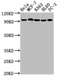
Positive WB detected in: Hela whole cell lysate, MCF-7 whole cell lysate, K562 whole cell lysate, HL60 whole cell lysate, PC-3 whole cell lysate
All lanes: TOP1 antibody at 1:2000
Secondary
Goat polyclonal to rabbit IgG at 1/50000 dilution
Predicted band size: 91 kDa
Observed band size: 91 kDa
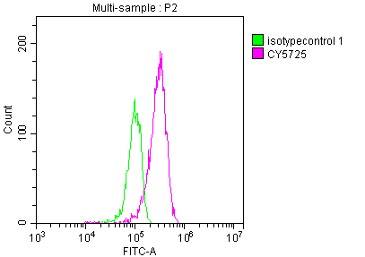
Overlay histogram showing HepG2 cells stained with CSB-RA792129A0HU (red line) at 1:50. The cells were fixed with 70% Ethylalcohol (18h) and then incubated in 10% normal goat serum to block non-specific protein-protein interactions followedby the antibody (1µg/1*106 cells) for 1 h at 4℃.The secondary antibody used was FITC-conjugated goat anti-rabbit IgG (H+L) at 1/200 dilution for 30min at 4℃. Control antibody (green line) was Rabbit IgG (1µg/1*106 cells) used under the same conditions. Acquisition of >10,000 events was performed.
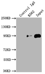
Immunoprecipitating TOP1 in K562 whole cell lysate
Lane 1: Rabbit control IgG instead of CSB-RA792129A0HU in K562 whole cell lysate. For western blotting,a HRP-conjugated Protein G antibody was used as the secondary antibody (1/2000)
Lane 2: CSB-RA792129A0HU(2µg)+ K562 whole cell lysate(500µg)
Lane 3: K562 whole cell lysate (10µg)
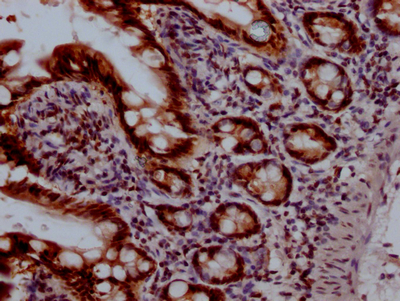
IHC image of CSB-RA792129A0HU diluted at 1:100 and staining in paraffin-embedded human small intestine tissue performed on a Leica BondTM system. After dewaxing and hydration, antigen retrieval was mediated by high pressure in a citrate buffer (pH 6.0). Section was blocked with 10% normal goat serum 30min at RT. Then primary antibody (1% BSA) was incubated at 4℃ overnight. The primary is detected by a Goat anti-rabbit IgG polymer labeled by HRP and visualized using 0.05% DAB.
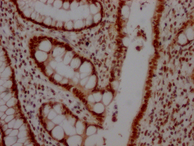
IHC image of CSB-RA792129A0HU diluted at 1:100 and staining in paraffin-embedded human colon cancer performed on a Leica BondTM system. After dewaxing and hydration, antigen retrieval was mediated by high pressure in a citrate buffer (pH 6.0). Section was blocked with 10% normal goat serum 30min at RT. Then primary antibody (1% BSA) was incubated at 4℃ overnight. The primary is detected by a Goat anti-rabbit IgG polymer labeled by HRP and visualized using 0.05% DAB.
TOP1 Antibodies
TOP1 for Homo sapiens (Human)
| 产品货号 | 产品名称 | 种属反应性 | 应用类型 |
|---|---|---|---|
| CSB-PA024056GA01HU | TOP1 Antibody | Human,Mouse,Rat | ELISA,WB |
| CSB-PA050283 | TOP1 Antibody | Human,Mouse,Rat | WB, ELISA |
| CSB-PA024056LA01HU | TOP1 Antibody | Human | ELISA, IF |
| CSB-PA024056LB01HU | TOP1 Antibody, HRP conjugated | Human | ELISA |
| CSB-PA024056LC01HU | TOP1 Antibody, FITC conjugated | Human | |
| CSB-PA024056LD01HU | TOP1 Antibody, Biotin conjugated | Human | ELISA |
| CSB-RA792129A0HU | TOP1 Recombinant Monoclonal Antibody | Human | ELISA, WB, IHC, FC, IP |
TOP1 Proteins
TOP1 Proteins for Homo sapiens (Human)
| 产品货号 | 产品名称 | 来源 |
|---|---|---|
| CSB-YP024056HU CSB-EP024056HU CSB-BP024056HU CSB-MP024056HU CSB-EP024056HU-B |
Recombinant Human DNA topoisomerase 1 (TOP1) | Yeast E.coli Baculovirus Mammalian cell In Vivo Biotinylation in E.coli |
| CSB-BP024056HU1 | Recombinant Human DNA topoisomerase 1 (TOP1), partial | Baculovirus |
| CSB-BP024056HU1c7 | Recombinant Human DNA topoisomerase 1 (TOP1), partial | Baculovirus |


